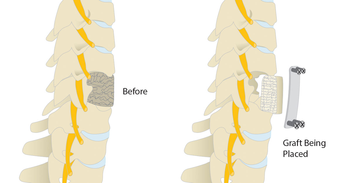
Cervical Corpectomy

Cervical Corpectomy
Anterior Cervical Corpectomy is a surgical procedure in which the vertebral bone and the intervertebral disc material is removed. The procedure is performed to relieve pain caused by stress on the spinal cord and spinal nerves in the neck.
This surgery involves accessing the cervical spine from the front. Due to the amount of vertebral bone or disc material that has to be removed to relieve pressure on the spinal cord or nerves, spinal fusion is typically needed.
Who needs Cervical Corpectomy?
Anterior Cervical Corpectomy is suggested for patients who have nerve compression in the cervical spine. The nerve compression in the cervical spine causes neck pain, numbness and weakness in the hands, arms, and shoulders.
This procedure is also suggested for patients who show symptoms such as pins and needles, tingling, or weakness in the hands and arms. More serious symptoms can be stumbling, loss of balance, and a loss of control of bowel and bladder.
Cervical Corpectomy is recommended only for patients who have gone through conservative treatment but these treatments failed.
What are the steps involved in Cervical Corpectomy?
Following steps are involved in the Anterior Cervical Corpectomy:
Incision
This surgical procedure approaches the neck from front. The surgeon cleans the skin and makes a small vertical incision in the front of the neck. The skin and neck tissues are gently retracted to access the cervical spine.
Removal of disc and vertebra
The surgeon removes the damaged disc between the upper and lower vertebrae. The diseased or damaged vertebra (or vertebrae) is also removed to relieve the pressure on the spinal nerves and spinal cord.
Placing bone graft
Then the surgeon cleans the space and prepares it for a bone graft or bone graft substitute. The bone graft is then placed between the adjacent vertebrae. The bone graft can be an autograft bone which is taken from the patient's own hip, or it can be an allograft bone taken from the bone bank.
Implanting metal plate
Ones the bone graft is fitted in its place, the surgeon screws a metal plate over the area. The screws and metal plate hold the bones in place while the vertebrae heal. When the vertebrae are in the process of healing, the bone graft knits with the upper and lower vertebrae to form a new bone mass called fusion.
Closure
The wound is then closed with a few stitches or medical glue. The bandage is applied after cleaning the wound.
After the Surgery
Generally the patients have to stay in the hospital for about 24 hours. In most of the cases they do not need to wear a cervical collar. Many patients will feel immediate relieve of pain and improvement of their symptoms. In some cases the patients may experience a sore throat or some hoarseness for a few days after the surgery. Usually the patients discontinue painkillers and resume their normal activities within a few weeks after the surgery.
